
Why Kir7.1 matters in Leber congenital amaurosis
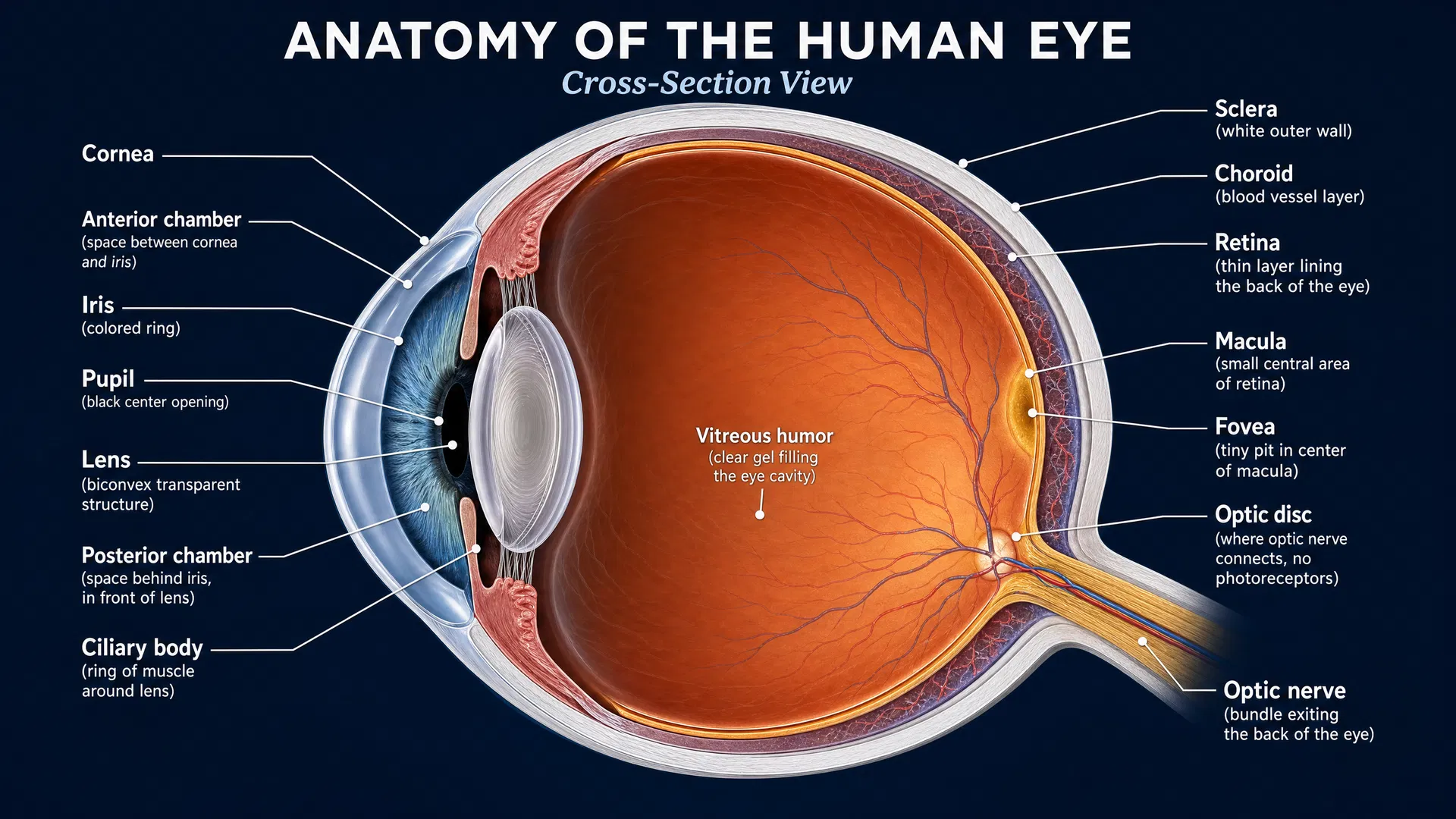
Leber congenital amaurosis (LCA) is a group of inherited retinal diseases that causes severe vision loss beginning at birth or in early childhood. Different genetic changes can lead to LCA, and some affect the retinal pigment epithelium (RPE)—a layer of cells that supports the light-sensing photoreceptors of the retina. One gene of interest is KCNJ13, which provides instructions for making the potassium channel Kir7.1.
When Kir7.1 function is disrupted in the RPE, it has been associated with retinal conditions including LCA, retinitis pigmentosa, and snowflake vitreoretinal degeneration. Understanding exactly how this channel is built, how it works, and how it can be controlled is therefore important for the broader effort to understand—and eventually treat—KCNJ13-related retinal disease.
A 2026 study in Nature Communications provides a detailed look at human Kir7.1. Although the work also examines the channel in brain circuits involved in appetite and body weight, its structural findings create valuable foundations for research into Kir7.1-associated eye disease.
A potassium channel with an important retinal role
Ion channels are protein gateways in cell membranes. They control the movement of charged particles, or ions, into and out of cells. Kir7.1 is an inwardly rectifying potassium channel, meaning that it helps regulate potassium movement across the cell membrane in a particular direction depending on electrical conditions.
In the RPE, healthy ion movement is part of the support system that helps maintain the retinal environment. When a channel such as Kir7.1 does not work properly, that support may be compromised. Over time, this can contribute to dysfunction of the photoreceptors that enable sight.
The new study used cryo-electron microscopy, a powerful imaging approach that can reveal the three-dimensional shapes of proteins. By determining structures of human Kir7.1, the researchers supplied a molecular-level view of the channel. Structural information is especially useful because disease-causing genetic variants may alter a channel’s shape, stability, location in the cell membrane, or ability to open and close.
Revealing how a small molecule blocks Kir7.1
The investigators also identified the structural basis for Kir7.1 blockade by ML418, a small molecule that inhibits the channel. In simple terms, the study shows how this compound can interact with Kir7.1 in a way that prevents normal channel activity.
This finding is not a treatment for LCA. In fact, because loss of Kir7.1 function is associated with retinal disease, blocking the channel would not be an obvious strategy for restoring Kir7.1 activity in the eye. However, knowing where and how a molecule binds is still highly informative. It gives researchers a map for designing future compounds and for testing how particular genetic changes might affect the channel’s behavior or its response to medicines.
The study also demonstrated that blocking Kir7.1 affects neurons that express the melanocortin-4 receptor (MC4R) in the paraventricular nucleus of the hypothalamus, a brain region involved in energy balance. Kir7.1 inhibition depolarized these neurons in brain slices and activated them in living animals. This was associated with reduced food intake and weight loss. These experiments emphasize that Kir7.1 has important functions beyond the eye and that treatments targeting the channel would need to consider effects in other tissues.
An unusual partnership with MC4R
A particularly intriguing aspect of the research is Kir7.1’s relationship with MC4R. MC4R is a G protein-coupled receptor (GPCR), a large family of receptors that typically relay signals through intermediary cellular pathways. Previous studies suggested that MC4R may regulate Kir7.1 more directly and without the usual G protein signaling route.
In this publication, the researchers found that Kir7.1 and MC4R are closely associated at the plasma membrane, the outer boundary of cells. They also developed and characterized a fusion construct joining MC4R and Kir7.1. This engineered research tool may make it easier to study their interaction in controlled experiments.
For families affected by LCA, this does not mean that MC4R is already a proven driver of KCNJ13-related retinal disease. Rather, it expands the scientific toolkit for investigating how Kir7.1 is regulated and whether its activity can be modified in precise ways.
What this could mean for future treatment research
Current work on inherited retinal diseases increasingly depends on understanding both the disease gene and the biology of the protein it encodes. For KCNJ13-related LCA, gene-based approaches may ultimately aim to address the underlying genetic cause. At the same time, detailed protein structures can support complementary strategies, such as identifying compounds that stabilize a faulty channel, improve its function, or help distinguish between variants with different biological effects.
The Kir7.1 structures and ML418 binding information reported in this study provide a starting framework for these efforts. They may also help researchers create better laboratory models for evaluating disease-associated KCNJ13 variants.
Looking ahead
This study does not deliver an immediate therapy for LCA, but it strengthens the foundation on which future therapies can be built. By defining human Kir7.1 structurally, explaining how a channel-blocking molecule acts, and creating new tools to examine its association with MC4R, the researchers have opened additional paths for investigation.
For the LCA community, progress often comes through this kind of stepwise research: connecting genetic findings to protein function, understanding the consequences in retinal cells, and using that knowledge to develop targeted interventions. Kir7.1 is now becoming a clearer molecular target—and that clarity is essential for the next generation of inherited retinal disease research.