Why measuring change matters in BVMD
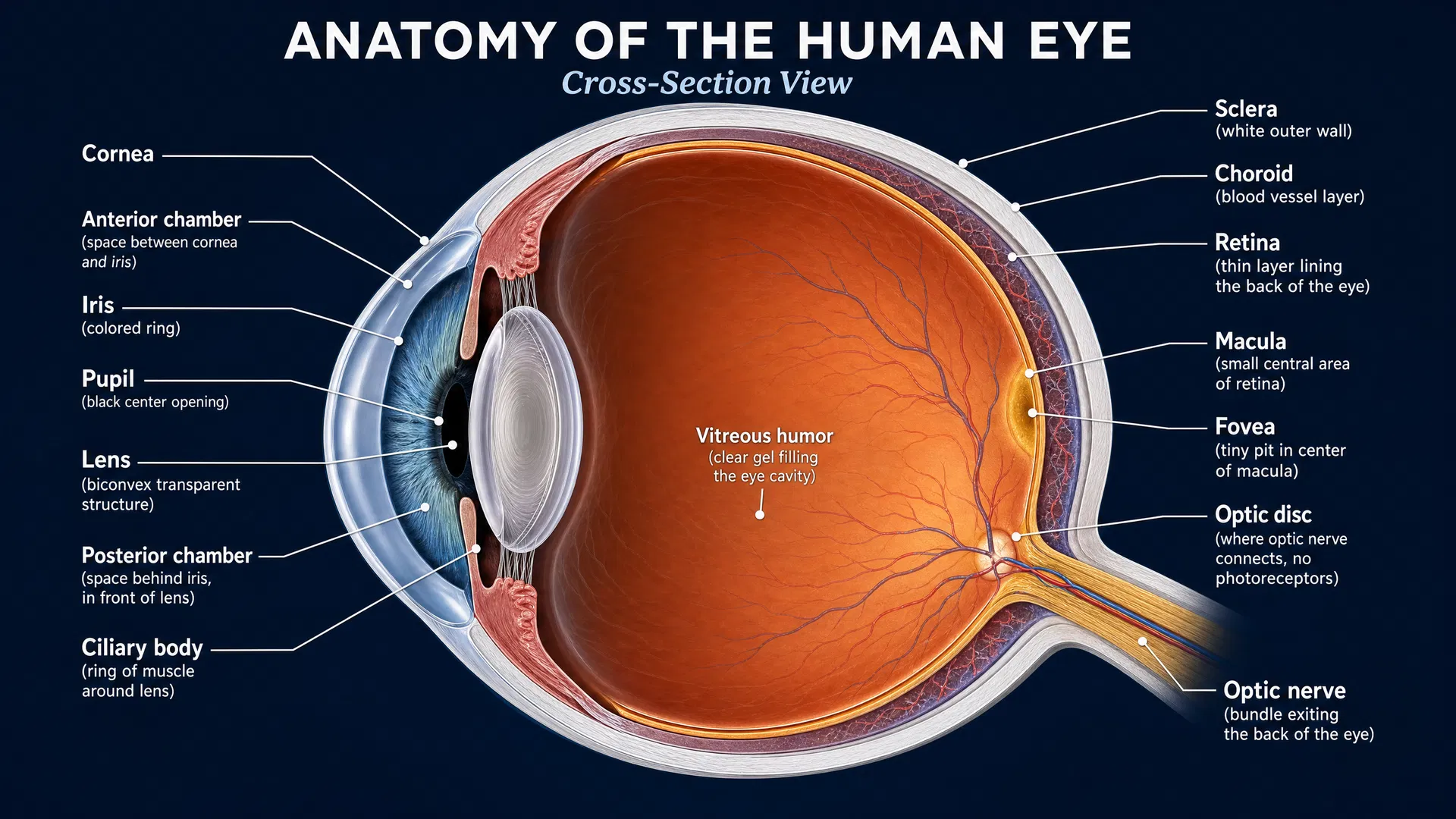
Best vitelliform macular dystrophy (BVMD), often called Best disease, is an inherited retinal condition that affects the macula—the central part of the retina used for detailed tasks such as reading, recognizing faces, and seeing fine detail. For people living with BVMD and their families, an important question is not only how vision is functioning today, but how it changes over time.
Answering that question is also essential for treatment development. Clinical trials need outcome measures that can reliably detect whether a therapy is slowing, stopping, or improving disease-related changes. In slowly progressive conditions such as BVMD, commonly used averages may appear stable even when small areas of retinal function are declining. A 2026 natural-history study explored whether microperimetry could reveal these subtle changes and help prepare for future clinical trials, including gene therapy studies.
What is microperimetry?
Microperimetry is a retinal function test that measures how sensitive different locations in the macula are to dim spots of light. Unlike a standard eye chart, which provides a broad measure of central visual acuity, microperimetry maps retinal sensitivity point by point.
In this study, participants underwent mesopic microperimetry, meaning testing under moderate, everyday-light conditions. Researchers used a 68-point grid covering the central 10 degrees of the visual field—the portion most relevant to fine central vision. Each test location was categorized using normative data:
- Normal: sensitivity was within the expected range.
- Relative scotoma: sensitivity was reduced, but some light detection remained.
- Deep scotoma: sensitivity was extremely low, below 1 decibel (dB).
This approach allowed the investigators to look beyond a single average score and examine where retinal sensitivity was changing.
The study: a window into BVMD’s natural history
The study included 23 eyes from 13 people with clinical BVMD enrolled in the IRDs-OSR natural-history cohort. Participants had a median baseline age of 29.4 years. On average, each eye was tested three times over a median follow-up period of 4.4 years.
At the beginning of the study, the median macular sensitivity across the central 10 degrees was 21.4 dB. Of all measured test points, 44% were classified as normal, 54% as relative scotomas, and 2% as deep scotomas. In other words, reduced sensitivity was already present at many tested locations, while profound loss was comparatively uncommon.
When the researchers averaged sensitivity across all 68 points, they found no statistically significant decline over time. The mean rate of change was minus 0.16 dB per year. This finding reinforces that, at a broad level, macular sensitivity in BVMD may appear relatively stable over several years.
Looking at normal areas revealed a clearer signal
The most important finding emerged when the team focused only on points that were normal at baseline. These areas showed a small but statistically significant decline in sensitivity, averaging minus 0.36 dB per year.
The decline was greatest in the central 5 degrees of the macula, where sensitivity fell by an average of minus 0.56 dB per year. This central region is particularly important because it supports the sharpest, most detailed vision.
The researchers also tracked changes in the number of deep scotoma points. On average, deep scotomas increased by 0.22 points per year—roughly one additional deep scotoma point every five years. Counts of normal points and relative scotoma points did not change significantly during the study period.
Together, these results show why averaging all retinal locations can obscure meaningful progression. Areas that already have reduced sensitivity may have limited room to decline further, while initially healthy-appearing areas can provide a more sensitive measure of early functional change.
Implications for clinical trials and future treatments
The study provides valuable information for designing BVMD clinical trials. If a trial relies only on overall average macular sensitivity, the slow progression of BVMD could make it difficult to detect a treatment effect over a practical trial timeframe.
By contrast, measuring sensitivity specifically at locations that are normal when a study begins may offer a more responsive outcome measure. Tracking these points could help investigators determine whether an intervention preserves retinal function that is still present. The central 5-degree region may be especially informative because it showed the steepest decline in this cohort.
This is particularly relevant to gene therapy research. Treatments intended to protect or restore retinal cells may be most effective before severe functional loss occurs. An outcome measure that captures change in relatively preserved retinal areas could therefore be important for evaluating whether a therapy is helping maintain central macular function.
A foundation for the next stage of BVMD research
This natural-history study highlights both the slow pace of BVMD progression and the value of precise functional testing. For patients and families, the findings offer a clearer picture of why changes may be difficult to notice from year to year, even while subtle retinal changes are occurring.
For researchers, the work identifies a practical microperimetry-based strategy for future trials: focus on retinal locations with normal sensitivity at baseline rather than relying solely on a whole-macula average. As treatment research advances, well-chosen outcome measures will be central to turning promising therapies into trials that can meaningfully assess protection of vision in BVMD.