
A finding that expands the genetic picture of cone-rod dystrophy
Cone-rod dystrophy is an inherited retinal disease in which cone photoreceptors—the cells supporting central, detailed, and color vision—are affected prominently, often followed by rod photoreceptors that support vision in dim light. Because many different genes can contribute to similar retinal symptoms, identifying the underlying genetic cause can be challenging for patients and families.
A 2026 study published in Molecular Genetics & Genomic Medicine reports two previously unreported variants in PCDH15 in a Chinese family affected by autosomal recessive cone-rod dystrophy. The findings provide preliminary evidence that changes in this gene may be associated with cone-rod dystrophy and broaden the range of genetic findings that researchers should consider when investigating this condition.
What the researchers found
The study focused on a 42-year-old woman with cone-rod dystrophy and evaluated members of her family. The research team combined detailed eye examinations with genetic testing, including whole-exome sequencing, a method that examines the protein-coding portions of many genes at once. They then used Sanger sequencing—a targeted DNA testing method—to confirm the candidate variants and study how they were inherited within the family.
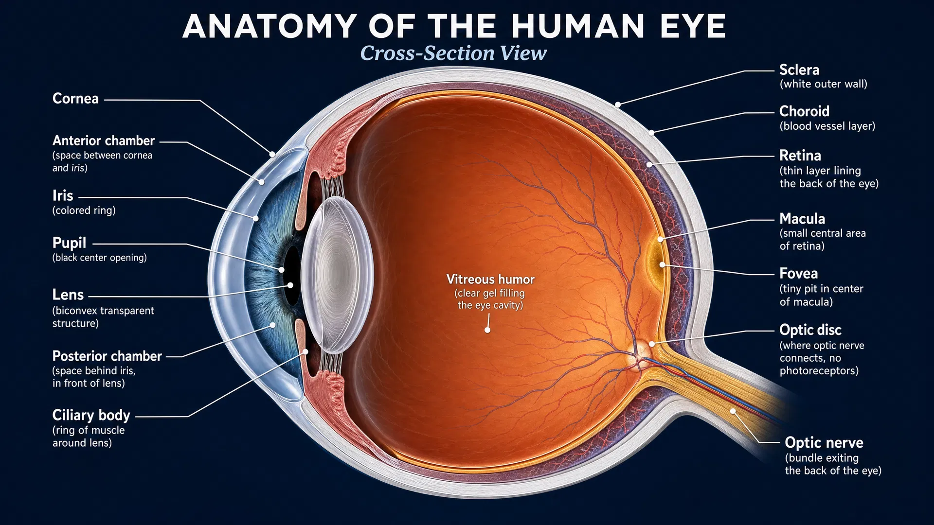
The participant’s retinal examinations showed changes consistent with retinal degeneration in both eyes. On ophthalmoscopic examination, the researchers observed pigmentary changes and areas of atrophy, or tissue loss, in the posterior pole and around the optic nerve head.
Optical coherence tomography (OCT), which provides cross-sectional images of the retina, showed atrophy of the outer retinal layers. These layers contain photoreceptors and supporting structures essential for vision. OCT also showed disruption of the ellipsoid zone at the posterior pole. The ellipsoid zone is an imaging feature associated with photoreceptor integrity, so its disruption is a marker of damage to these light-sensing cells.
Functional testing supported the structural findings. Multifocal electroretinography (mfERG), which measures electrical responses from different retinal regions, found reduced responses in the central retina. Full-field electroretinography (ffERG), which measures retinal activity across the eye, showed a more substantial reduction in cone-driven responses under light-adapted, or photopic, conditions. This pattern aligns with cone-predominant retinal dysfunction.
Two novel variants in PCDH15
The genetic analysis identified two different PCDH15 variants in the affected individual: c.4903_4906del (p.Glu1635Lysfs*4) and c.3470C>A (p.Ala1157Glu). Having two altered copies of a gene, with one inherited from each parent, is known as compound heterozygosity. This inheritance pattern is consistent with autosomal recessive disease.
One variant is a small deletion predicted to shift the gene’s reading frame and introduce an early stop signal, which could substantially alter the resulting protein. The other changes a single amino acid, replacing alanine with glutamic acid at position 1157. Family co-segregation analysis provided supportive evidence that these variants tracked with the disease in the pedigree.
The investigators also examined the PCDH15 protein across species and found that it is highly conserved, meaning important portions of the protein have been maintained through evolution. Structural modeling further predicted that the identified variants could alter protein structure. Together, these observations support the possibility that the variants are relevant to the observed retinal phenotype, although the authors describe the evidence as preliminary.
Why this matters for families
For people with cone-rod dystrophy, a genetic diagnosis can help explain why retinal disease has developed and clarify its inheritance pattern within a family. This report also illustrates the value of pairing genetic testing with clinical measures such as retinal imaging and electroretinography. Genetic results are most informative when considered alongside the individual’s eye findings and family history.
Importantly, this study is described as the first report associating compound heterozygous PCDH15 variants with cone-rod dystrophy. A single-family report does not establish the full range of PCDH15-related retinal disease, but it gives researchers and diagnostic laboratories a new gene–phenotype association to investigate in additional families.
Implications for treatment research
The publication does not test a treatment or report a therapy directed at PCDH15. Its contribution is instead foundational: defining genetic causes and associated retinal features helps build the knowledge needed for future therapeutic research.
As research advances, well-characterized families can help scientists understand how particular gene changes affect photoreceptors, why disease severity may vary, and which clinical measures best track retinal change over time. OCT, mfERG, and ffERG findings from reports such as this one may also be useful in shaping future natural-history studies.
Looking ahead
This study adds an important early piece to the cone-rod dystrophy research landscape. Replication in more individuals and families will be essential to confirm the association between biallelic PCDH15 variants and this retinal condition. Further work can also examine how these variants affect PCDH15 function in the retina.
For now, the report reinforces a central lesson in inherited retinal disease research: each carefully studied family can expand understanding of the genes involved in retinal degeneration. As the genetic map of cone-rod dystrophy becomes more complete, it can strengthen diagnosis, family-based genetic evaluation, and the scientific groundwork for future treatment development.