Why better measurements matter
Malattia Leventinese, also called Doyne honeycomb retinal dystrophy, is a rare inherited retinal disease marked by the buildup of drusen—yellowish deposits beneath the retina—and progressive vision loss. Because its retinal changes resemble those seen in dry age-related macular degeneration (AMD), studying Malattia Leventinese can deepen understanding of both conditions. Yet its rarity also makes each clinical study especially important: trials need outcome measures that can reliably detect meaningful change over a practical period of time.
A 2026 multicenter natural-history study published in Investigative Ophthalmology & Visual Science followed 25 genetically confirmed participants for two years. The researchers examined which imaging and visual-function measures changed most clearly as disease progressed. Their findings identify several potential “surrogate endpoints”—measurements that may help researchers assess whether a future treatment is slowing retinal damage before large changes in everyday visual acuity become apparent.
Looking beneath the retina’s overall thickness
Participants underwent several forms of retinal assessment, including spectral-domain optical coherence tomography (SD-OCT), fundus photography, fundus autofluorescence, and microperimetry. SD-OCT creates cross-sectional images of the retina, allowing individual layers to be measured. Microperimetry tests light sensitivity at specific retinal locations, rather than relying only on a standard eye-chart result.
One of the study’s clearest messages is that total retinal thickness can be misleading in Malattia Leventinese. Over two years, the overall thickness of the retina remained stable. At first glance, that might suggest little progression. However, the layer-by-layer analysis showed substantial changes happening in opposite directions.
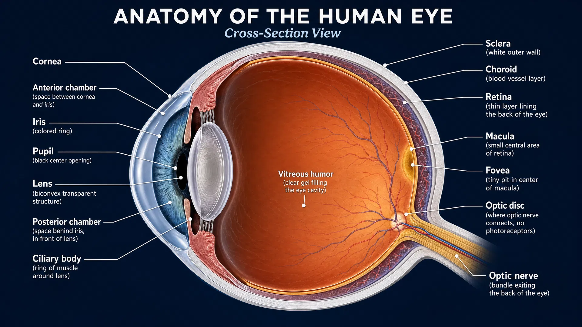
The retinal pigment epithelium and drusen complex (RPEDC) became thicker by an average of 2.24 micrometers per year. This reflects increasing drusen-related material in and beneath the retinal pigment epithelium, a supportive cell layer essential for photoreceptor health. At the same time, important outer retinal layers became thinner:
- Photoreceptor inner segments declined by 0.70 micrometers per year.
- The outer nuclear layer declined by 1.95 micrometers per year.
- Choroidal thickness declined by 10.83 micrometers per year.
The outer nuclear layer contains the cell bodies of photoreceptors, the light-sensing cells that support vision. The inner segments are metabolically active portions of these cells. Their thinning provides structural evidence of photoreceptor degeneration, even when total retinal thickness appears unchanged because growing drusen offset the loss of retinal tissue.
Night-vision testing detected decline earlier
The functional findings were equally important. The study used mesopic microperimetry, which tests sensitivity under moderate lighting, and two-color scotopic microperimetry, which tests sensitivity in dark-adapted conditions. Only scotopic sensitivity showed a significant decline over time: an average loss of 0.40 decibels per year at 505 nm and 0.37 decibels per year at 627 nm. Mesopic sensitivity remained stable during the study period.
In accessible terms, the study suggests that the retina’s ability to detect dim light may worsen before a measurable decline is captured under brighter, everyday testing conditions. This is consistent with the experience that difficulties in low-light environments can be an important feature of retinal disease, even when conventional visual measures do not change substantially.
The researchers also found that functional decline was greatest in earlier disease stages, around the onset of drusen accumulation, and that scotopic function decreased before age 55. In contrast, mesopic testing may be more useful for following later disease stages.
What this means for treatment development
Natural-history studies do not test a treatment, but they create the foundation for treatment trials. To determine whether an investigational therapy works, researchers need measures that are sensitive, repeatable, and closely connected to disease progression.
This study supports outer nuclear layer thickness and photoreceptor inner-segment thickness as particularly useful structural endpoints. These measures directly track layers affected by photoreceptor degeneration. It also supports scotopic microperimetry as an early functional endpoint, especially when researchers need to detect changes that may occur before mesopic sensitivity declines.
The findings also underscore why relying on total retinal thickness alone could miss meaningful progression in Malattia Leventinese. A treatment that reduces drusen-related thickening but does not preserve photoreceptors, for example, would require careful interpretation using multiple imaging and functional measures. Combining detailed OCT layer analysis with dark-adapted sensitivity testing may provide a more complete picture of whether a therapy is protecting retinal structure and function.
A clearer path for future studies
With 25 participants, this was a small but valuable study in a rare genetic condition. Its two-year follow-up and multimodal approach offer practical evidence for selecting endpoints in ongoing and future research. The results may also help investigators design studies that enroll people at stages when change can be detected most efficiently.
For patients and families, the study reinforces that disease progression can be measurable even when broad retinal thickness or standard daytime visual testing seems stable. As research moves forward, precise layer-specific imaging and low-light functional testing may help bring Malattia Leventinese trials closer to answering the questions that matter most: whether a therapy preserves photoreceptors, protects vision, and changes the course of disease.