
Why this study matters
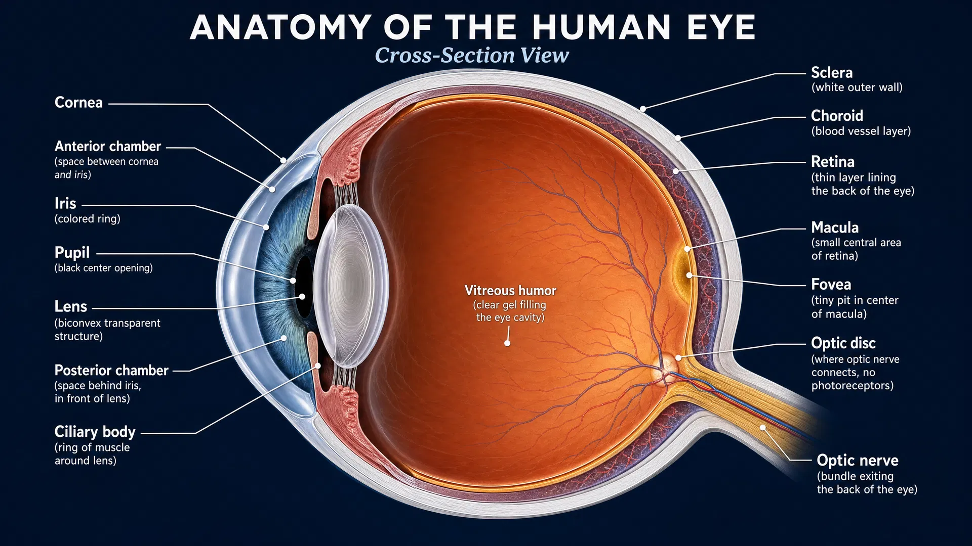
Choroideremia (CHM) is an inherited retinal disease caused by disease-causing variants in the CHM gene. It progressively affects the retina and can narrow the visual field over time, often while central reading vision remains relatively preserved until later stages. For people living with CHM and their families, an important research question is not only whether vision changes, but also *which tests can measure those changes accurately enough to evaluate potential treatments.
This 48-month prospective natural history study, published in Ophthalmology Science, provides one of the most detailed looks at how structural retinal changes, visual-field performance, and patient-reported visual function progress over time. Its results help identify practical, reliable endpoints for future CHM clinical trials.
Study design: following CHM over time
The study enrolled 57 male participants, ages 14 to 70, across five centers in the United States and Canada. Every participant had a clinical diagnosis of CHM and a disease-causing CHM variant. Participants also had relatively preserved central retinal structure and best-corrected visual acuity of at least 20/200 at enrollment.
Testing occurred every six months for the first 24 months. Participants who completed that period were invited to continue follow-up visits through month 48. Researchers assessed several aspects of disease progression:
- Best-corrected visual acuity (BCVA): the ability to read standardized eye-chart letters
- Static and kinetic visual fields: tests of sensitivity to light or the extent of the remaining visual field
- Preserved fundus autofluorescence (PFAF): imaging that identifies areas of retinal tissue still showing autofluorescence
- Ellipsoid zone (EZ) width on OCT: a scan-based measure of a retinal layer associated with photoreceptor integrity
- Visual Function Questionnaire-25 (VFQ-25): a questionnaire capturing how vision affects daily life
Central visual acuity changed little, but other measures revealed progression
Over the study period, visual acuity showed very little average annual change: an estimated loss of 0.17 letters per year in right eyes and 0.16 letters per year in left eyes. Neither result was statistically significant.
This is an important finding. In CHM, standard visual acuity testing may remain stable even as the disease continues to reduce the functioning retina outside the center of vision. A person may still read eye-chart letters well while experiencing worsening peripheral vision, dim-light difficulties, or limitations in daily activities. As a result, visual acuity alone may not be the most sensitive endpoint for studies involving participants with preserved central vision.
Visual-field tests showed meaningful functional decline
The study found measurable change in visual fields, although the usefulness of kinetic testing depended on the test target used. The largest target, called size 14e, showed the fastest decline, but many participants could not perform that test reliably. With smaller III4e and V4e targets, annual changes ranged from 13.7% to 20.4%.
Static perimetry, which measures sensitivity to light at multiple locations, also detected consistent progression. Average sensitivity declined by 8.1% per year in right eyes and 9.6% per year in left eyes. These findings indicate that visual-field measures can capture the functional consequences of retinal degeneration, even when central acuity remains stable.
Participants’ own reports supported this picture. VFQ-25 composite scores declined by an average of 4.2 points during the first 24 months. The researchers noted that this degree of change is clinically meaningful to participants, emphasizing that disease progression can affect day-to-day vision before a substantial change appears on an eye chart.
Retinal imaging identified a precise structural endpoint
PFAF area declined by approximately 10% each year: 10.1% in right eyes and 9.7% in left eyes. The researchers concluded that PFAF area has advantages over measuring EZ area as a CHM clinical-trial endpoint because it is easier to measure and can be measured more precisely.
OCT imaging also documented loss of the ellipsoid zone. The vertical dimension declined faster than the horizontal dimension, with annual decreases of about 9.9% to 10.4% vertically and 5.9% to 7.0% horizontally. This difference suggests that retinal change in CHM may not occur uniformly in every direction.
Crucially, several functional visual-field measures correlated strongly with PFAF area. That relationship strengthens confidence that PFAF is not simply an imaging change: it reflects a structural measure that tracks with visual function.
What this means for treatment development
Natural history studies establish the expected course of disease in the absence of an investigational intervention. In future CHM treatment studies, researchers can compare changes in treated participants with these expected rates of decline.
The results suggest that preserving PFAF area may be a particularly useful way to assess whether an approach slows retinal degeneration. An average annual PFAF decrease of about 10% provides a concrete benchmark for trial planning. Visual-field testing and patient-reported outcomes may add essential evidence about whether structural preservation is accompanied by meaningful functional benefit.
A stronger foundation for the next generation of CHM studies
This multicenter study shows why measuring CHM progression requires more than a standard visual-acuity chart. Imaging, visual fields, and participants’ reports of daily visual function each captured different parts of the disease experience.
By identifying PFAF area as a practical and precise structural endpoint—and linking it to functional visual-field measures—this research gives future CHM trials a stronger framework for detecting change. That framework can help researchers test whether emerging approaches preserve retinal tissue and, most importantly, maintain useful vision for longer.