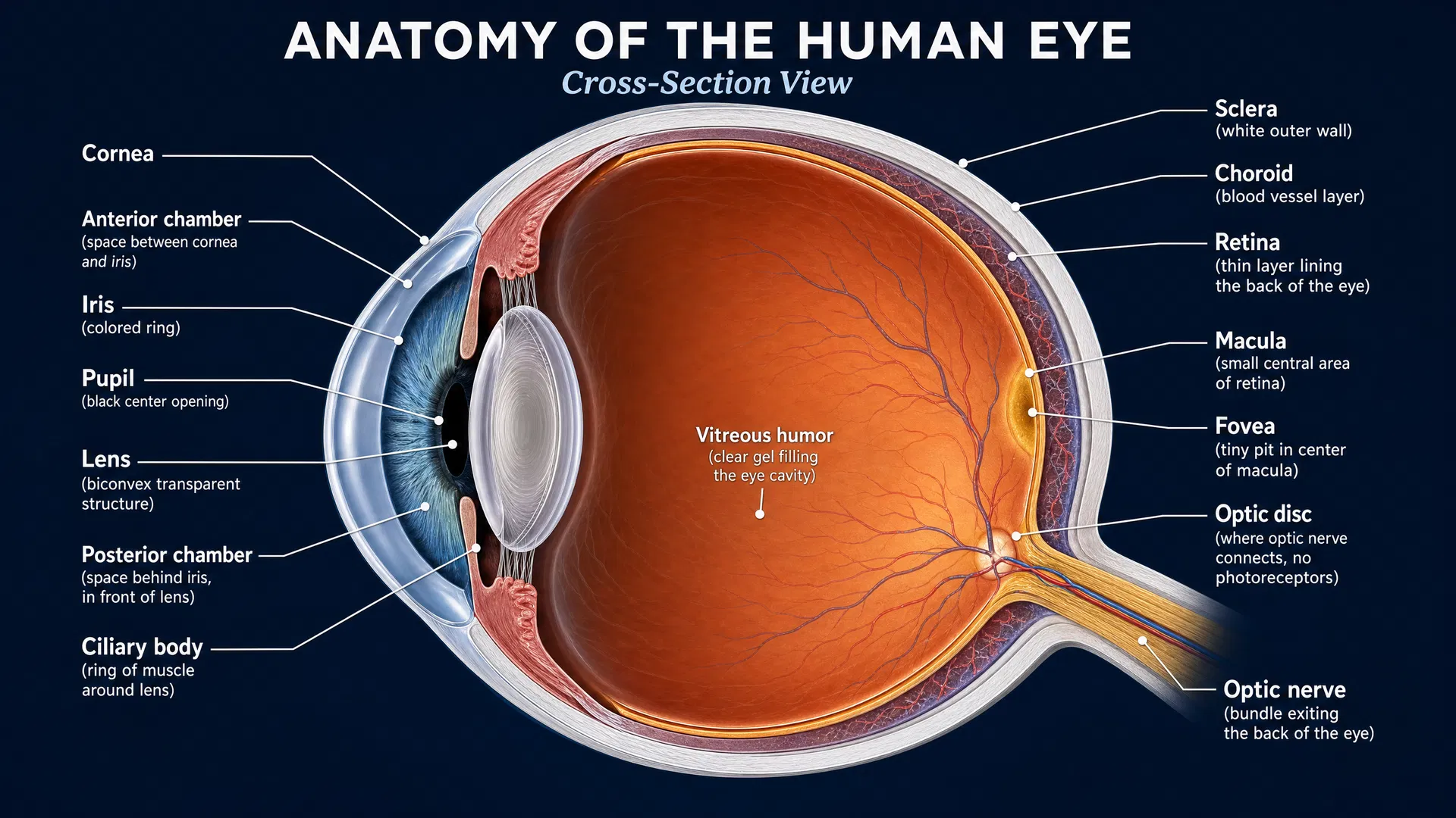
Why this finding matters
Retinitis pigmentosa (RP) is a group of inherited retinal diseases marked by progressive loss of light-sensitive cells in the retina. Because many genes can cause RP, identifying the underlying genetic explanation can be challenging—especially when a gene is usually associated with a very different, multisystem condition.
A 2026 study in the American Journal of Human Genetics reports that certain changes in IDUA can cause late-onset RP with few, or even no, recognizable features of mucopolysaccharidosis type I (MPS I). This expands the known range of IDUA-related disease and offers an important message for people and families affected by RP: a diagnosis may sometimes lie in genes traditionally linked to metabolic disorders, even when the expected systemic signs are absent.
IDUA and MPS I: the usual connection
Both copies of most genes are inherited—one from each parent. When a person has disease-causing changes in both copies of IDUA, the classic outcome is MPS I, a lysosomal storage disorder. Lysosomes are cellular structures that help break down and recycle materials. The IDUA gene provides instructions for the enzyme alpha-L-iduronidase, which is needed for this process.
In classic MPS I, little or no functional enzyme is available. This can lead to effects in multiple parts of the body. In contrast, the individuals described in this study had retinal disease consistent with RP, but a markedly milder overall pattern of disease.
The study’s key findings
Researchers identified 14 individuals from 12 families with RP who had disease-associated variants in both copies of IDUA. Their findings showed that IDUA-related disease can be retina-predominant or, based on assessments to date, apparently limited to the eye.
Most strikingly, seven individuals had no features consistent with MPS I after comprehensive systemic evaluation. At their most recent assessments, these individuals were between 46 and 73 years old. This is especially notable because it shows that an individual can reach later adulthood with IDUA-associated retinal degeneration without developing the recognizable multisystem picture of classic MPS I.
Testing of white blood cells in nine participants found reduced alpha-L-iduronidase activity in every person tested. However, activity was not completely absent. This residual enzyme function appears central to understanding why these individuals had milder disease and relative protection from systemic involvement.
“Hypomorphic” variants and residual enzyme activity
The study describes the participants’ IDUA genotypes as hypomorphic. In reader-friendly terms, this means the genetic changes reduce gene function rather than eliminating it entirely.
No participant had two IDUA variants expected to fully stop enzyme production or function. Most had one loss-of-function variant paired with a missense variant. A missense variant changes a single building block in the protein and can have effects ranging from minimal to severe.
The researchers used cell-based experiments to examine several missense variants. They measured enzyme activity and protein abundance, then calculated relative specific activity—a way to assess function while accounting for how much protein is present. Several variants retained only very low levels of function, typically around 0.5% to 1.5%. Even this small amount of residual activity may help explain why the disease was milder outside the retina.
One recurrent variant, c.298A>G (p.Arg100Gly), was particularly informative. Initial protein-focused testing did not show a direct effect on the protein or its catalytic activity. But long-read RNA analysis revealed that the variant predominantly disrupted RNA splicing, the process cells use to assemble a usable gene message. Most transcripts were abnormal and loss-of-function, while approximately 1% to 2% were correctly spliced. That small correctly spliced fraction provides a compelling explanation for the residual enzyme activity seen in affected individuals.
What this could mean for diagnosis and treatment research
For RP care, the study reinforces the value of looking beyond a strict divide between “retinal genes” and “metabolic genes.” When bi-allelic IDUA variants are found in someone with RP, genomic results can be strengthened by biochemical testing of alpha-L-iduronidase activity and, where needed, functional studies of individual variants.
This integrated approach matters because different variant types may affect the enzyme in different ways. Some may reduce protein abundance or processing, while others—such as p.Arg100Gly—may chiefly alter RNA splicing. Understanding the mechanism can clarify whether a variant truly contributes to disease and may help define which individuals have residual activity associated with systemic sparing.
The findings also establish an important foundation for future therapeutic research. A retina-predominant form of IDUA-associated disease creates a distinct group for studying how much enzyme activity is needed to protect tissues and why the retina may remain vulnerable despite low residual function. These questions are relevant to the development and evaluation of approaches designed to restore, increase, or preserve IDUA activity.
A changing view of inherited retinal disease
This study broadens the clinical spectrum of IDUA-associated disease from classic multisystem MPS I to late-onset RP that may be retina-predominant or apparently isolated. It also illustrates a broader lesson in inherited retinal disease research: genetic diagnoses become more powerful when DNA findings are considered alongside enzyme measurements, RNA studies, and laboratory functional testing.
As sequencing and functional tools continue to advance, researchers are likely to identify additional attenuated forms of metabolic disease among people initially diagnosed with isolated retinal degeneration. For families with RP, this progress may lead to more precise diagnoses, a clearer understanding of disease biology, and better-defined opportunities for future research.