Vitelliform Macular Dystrophy
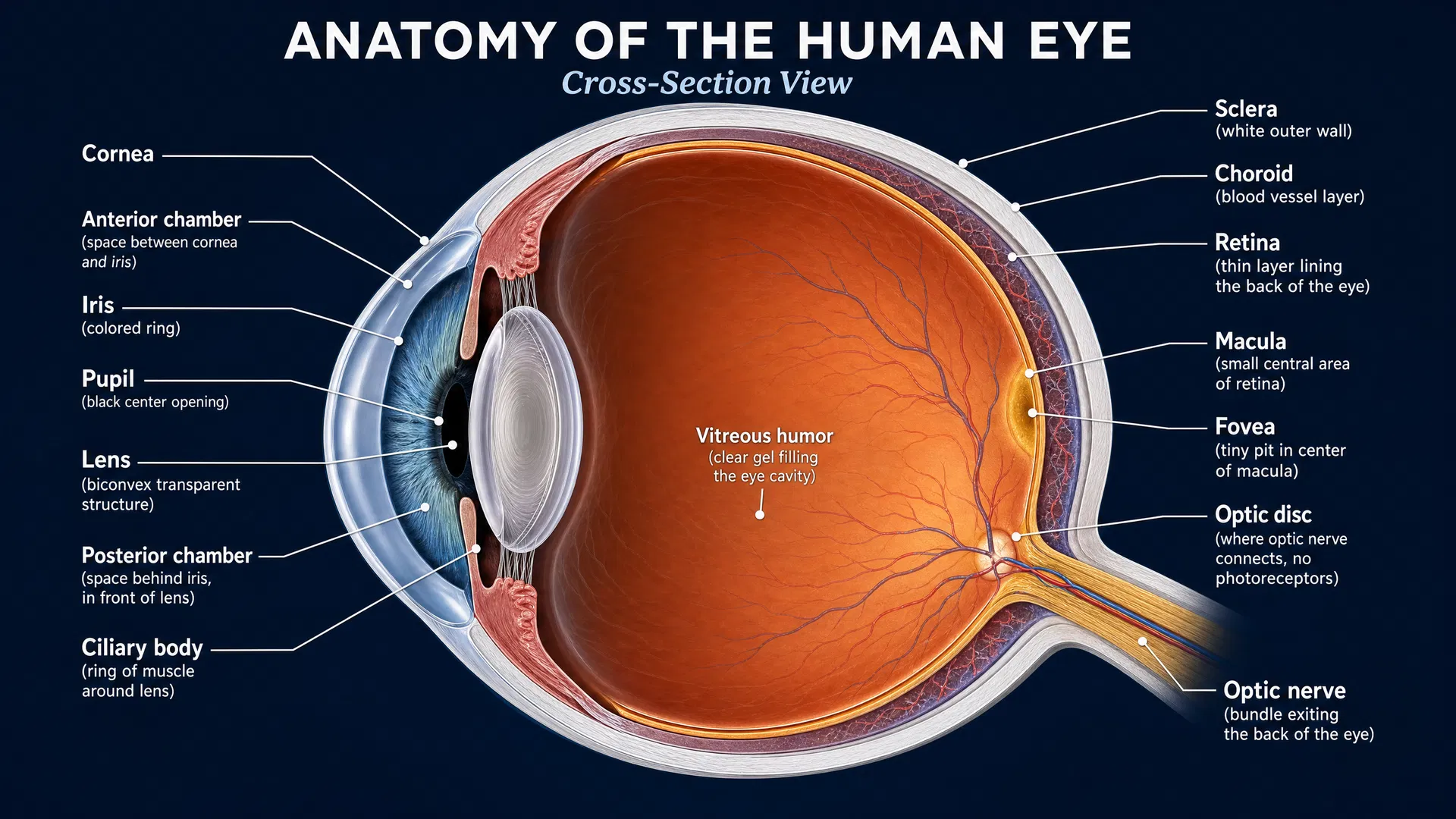
Adult-onset vitelliform macular dystrophy (AVMD) is a rare eye condition that affects the macula, the central part of the retina responsible for sharp, detailed vision. In this condition, a fatty yellow pigment called lipofuscin builds up underneath the macula, forming a lesion that can look like an egg yolk. This buildup can eventually damage the light-sensitive cells in the eye, leading to a gradual loss of central vision. Unlike some other eye diseases, AVMD usually begins in adulthood, typically between the ages of 40 and 60. Many people with AVMD may not notice any symptoms at first, or they might experience mild blurriness or a slight distortion in their vision, where straight lines appear wavy. The disease usually progresses very slowly, and most people maintain good enough vision to read and drive for many years. However, over time, the central vision can worsen, making it difficult to recognize faces or read small print. The condition generally does not affect side (peripheral) vision or the ability to see at night. While there is currently no cure for AVMD, regular eye exams are crucial to monitor the condition and catch any complications early. In some cases, abnormal blood vessels can grow beneath the retina, causing rapid vision loss. If this happens, treatments like eye injections can be very effective at preserving sight. Patients are often advised to use an Amsler grid at home to check for sudden changes in their vision, maintain a healthy diet, wear sunglasses outdoors, and avoid smoking to help protect their remaining vision.
Condition category: Macular Dystrophy
Prevalence: 1 in 10,000
Inheritance patterns: Autosomal Dominant
Age of onset: Typically between the fourth and sixth decades of life (30 to 60 years), though it can range from 30 to 80 years.
Clinical overview: Note: Vitelliform Macular Dystrophy is the broader clinical term that encompasses Best Disease (Best Vitelliform Macular Dystrophy). See the Best Disease entry for the primary inherited form. Adult-onset vitelliform macular dystrophy (AOVMD) may be sporadic or associated with PRPH2/BEST1 mutations. Adult-onset vitelliform macular dystrophy (AVMD), also known as adult-onset foveomacular vitelliform dystrophy (AOFVD), is a rare, genetic macular dystrophy characterized by the accumulation of yellowish, egg yolk-like (vitelliform) material in the foveal or parafoveal region of the retina. It is classified within the broader group of pattern dystrophies of the retinal pigment epithelium (RPE). The condition typically manifests in adulthood, most commonly between the fourth and sixth decades of life, distinguishing it from the childhood-onset Best vitelliform macular dystrophy. Clinically, AVMD presents with bilateral, often asymmetric, solitary, round or oval subretinal lesions. Patients may be asymptomatic initially or present with mild visual disturbances such as blurred vision or metamorphopsia (distortion of vision). The disease generally progresses slowly, with most individuals maintaining relatively good central vision for many years. However, significant visual impairment can occur in later stages due to geographic atrophy of the RPE and photoreceptors or the development of complications like choroidal neovascularization (CNV). Accurate diagnosis of AVMD is clinically significant because it is frequently misdiagnosed as age-related macular degeneration (AMD) due to overlapping phenotypic features. Differentiating AVMD from AMD is crucial to avoid unnecessary treatments, such as anti-VEGF injections, unless CNV is present. The condition is genetically heterogeneous, with mutations in genes such as PRPH2, BEST1, IMPG1, and IMPG2 identified in a minority of cases, while most cases appear sporadic. The relevant OMIM numbers for AVMD and its genetic variants include 153840, 608161, 616151, and 616152, and its Orphanet number is ORPHA:99000.
Patient and family guide: Adult-onset vitelliform macular dystrophy (AVMD) is a rare eye condition that affects the macula, the central part of the retina responsible for sharp, detailed vision. In this condition, a fatty yellow pigment called lipofuscin builds up underneath the macula, forming a lesion that can look like an egg yolk. This buildup can eventually damage the light-sensitive cells in the eye, leading to a gradual loss of central vision. Unlike some other eye diseases, AVMD usually begins in adulthood, typically between the ages of 40 and 60. Many people with AVMD may not notice any symptoms at first, or they might experience mild blurriness or a slight distortion in their vision, where straight lines appear wavy. The disease usually progresses very slowly, and most people maintain good enough vision to read and drive for many years. However, over time, the central vision can worsen, making it difficult to recognize faces or read small print. The condition generally does not affect side (peripheral) vision or the ability to see at night. While there is currently no cure for AVMD, regular eye exams are crucial to monitor the condition and catch any complications early. In some cases, abnormal blood vessels can grow beneath the retina, causing rapid vision loss. If this happens, treatments like eye injections can be very effective at preserving sight. Patients are often advised to use an Amsler grid at home to check for sudden changes in their vision, maintain a healthy diet, wear sunglasses outdoors, and avoid smoking to help protect their remaining vision.
Symptoms and clinical features: In the early stages of Adult-Onset Vitelliform Macular Dystrophy (AVMD), patients are often completely asymptomatic. The condition may be discovered incidentally during a routine eye examination. When symptoms do occur early on, they are typically mild and may include slight blurring of central vision or a small central or paracentral scotoma (blind spot). Patients might also notice mild metamorphopsia, where straight lines appear slightly wavy or distorted. Visual acuity in the early stages usually ranges from 20/20 to 20/50. As the disease progresses to the intermediate stages, the vitelliform lesion may undergo changes, sometimes breaking up or resorbing (the vitelliruptive stage). During this time, the blurring of central vision and metamorphopsia may become more noticeable and slowly worsen. Patients may begin to experience more difficulty with tasks requiring fine visual detail, such as reading small print or recognizing faces from a distance. However, the decline in vision is generally slow and gradual. In the advanced stages of AVMD, the vitelliform material often disappears, leaving behind an area of geographic atrophy (thinning and loss of tissue) in the macula. This leads to a more significant and permanent decrease in central visual acuity, sometimes dropping to 20/100 or worse. A major complication that can occur at any stage, but often leads to sudden and severe vision loss, is the development of choroidal neovascularization (CNV). If CNV occurs, patients may experience a rapid onset of severe metamorphopsia, a dense central scotoma, and significant visual impairment due to fluid leakage and bleeding under the retina.
Molecular pathology: The exact molecular pathophysiology of Adult-Onset Vitelliform Macular Dystrophy (AVMD) remains incompletely understood, but it involves the abnormal accumulation of lipofuscin and other material between the retinal pigment epithelium (RPE) and the photoreceptor layer. This accumulation is thought to result from disordered metabolism and impaired phagocytosis by the RPE cells, exacerbated by age, genetic predisposition, and environmental factors. The genes associated with AVMD provide insight into its molecular mechanisms. The PRPH2 gene encodes peripherin-2, a glycoprotein essential for the formation and stabilization of rod and cone outer segment discs. Mutations in PRPH2 may disrupt outer segment structure and renewal, leading to accumulation of debris. The BEST1 gene encodes bestrophin-1, a transmembrane protein in the RPE that functions as a calcium-activated chloride channel and regulates intracellular calcium signaling. BEST1 mutations impair these ionic channel functions, compromising the RPE-photoreceptor interface and leading to fluid and lipofuscin accumulation. The IMPG1 and IMPG2 genes encode interphotoreceptor matrix proteoglycans (SPACR and SPACRC, respectively) that are secreted into the extracellular matrix and play a role in retinal adhesion and structural integrity. Mutations in these genes disrupt the interphotoreceptor matrix, contributing to the separation of the RPE from the photoreceptors and the formation of vitelliform lesions. Over time, the accumulation of subretinal material and metabolic stress can lead to RPE hypertrophy, migration of pigment-laden macrophages, and eventual RPE and photoreceptor atrophy.
Genetics: Adult-onset vitelliform macular dystrophy (AVMD) is genetically heterogeneous. While early reports suggested an autosomal dominant inheritance pattern, it is now recognized that most cases are sporadic with no clear family history. When inherited, it typically follows an autosomal dominant pattern with variable expression and incomplete penetrance. Mutations in several genes have been associated with AVMD, including PRPH2, BEST1, IMPG1, and IMPG2. The PRPH2 gene (formerly RDS) is the most commonly identified, accounting for 2-18% of cases. BEST1 mutations, which are also responsible for Best disease, are found in a subset of patients, leading some to consider AVMD with BEST1 mutations as a late-onset, milder variant of Best disease. More recently, mutations in IMPG1 and IMPG2 have been identified in a small percentage (less than 8%) of AVMD patients. Despite these known genetic associations, the majority of AVMD patients do not have an identifiable mutation in these genes, suggesting that other genetic or environmental factors may contribute to the disease pathogenesis. Single nucleotide polymorphisms (SNPs) in genes like HTRA1 have also been investigated as potential risk factors.
Diagnostic evaluation: Diagnosis of Adult-Onset Vitelliform Macular Dystrophy (AVMD) relies on a comprehensive ophthalmologic examination. Fundus biomicroscopy typically reveals bilateral, asymmetric, solitary, round or oval subretinal lesions in the fovea or perifovea that are yellow and slightly elevated, often with a central pigmented spot. These lesions are generally 1/3 to 1 disc diameter in size. Fundus autofluorescence (FAF) imaging shows hyper-autofluorescence corresponding to the vitelliform lesions. Optical coherence tomography (OCT) is crucial for diagnosis and differentiation from other macular diseases. OCT reveals a vitelliform lesion located at the level of the retinal pigment epithelium (RPE) or between the RPE and photoreceptors, appearing as a dome-shaped hyper-reflective subretinal deposit. OCT is also essential for monitoring complications such as choroidal neovascularization (CNV) or subretinal fluid. Fluorescein angiography (FA) or OCT angiography (OCTA) may be used when CNV is suspected. Electrophysiological testing helps differentiate AVMD from Best disease. In AVMD, the electrooculogram (EOG) and full-field electroretinogram (ERG) typically show normal or subnormal results (normal Arden ratio), indicating localized macular pathology rather than generalized RPE dysfunction. Genetic testing for mutations in PRPH2, BEST1, IMPG1, and IMPG2 can support the diagnosis, though many cases are sporadic without identifiable mutations. Differential diagnosis includes Best vitelliform macular dystrophy (which typically has an earlier onset and abnormal EOG), age-related macular degeneration (AMD), Stargardt disease, central serous chorioretinopathy, and other pattern dystrophies. Differentiating AVMD from AMD is particularly important to avoid unnecessary treatments, as AVMD lesions can mimic drusen or pigment epithelial detachments seen in AMD.
Differential diagnosis: Differential diagnosis of adult-onset vitelliform macular dystrophy includes: (1) Best disease — earlier onset, larger lesion, markedly abnormal EOG, BEST1 mutations. (2) Pattern dystrophy — various pigment patterns, PRPH2 mutations. (3) Central serous chorioretinopathy — serous detachment, no vitelliform material. (4) Age-related macular degeneration — drusen, older onset, no vitelliform deposit. (5) Batten disease (CLN3) — vitelliform-like changes with neurodegeneration in children. (6) Paraneoplastic vitelliform maculopathy — associated with systemic malignancy, anti-retinal antibodies.
Natural history: Adult-onset vitelliform macular dystrophy typically presents between the fourth and sixth decades of life. The disease follows a slow, progressive course characterized by the evolution of the vitelliform lesion. Initially, the lesion appears as a yellow, dome-shaped subretinal deposit. Over years to decades, this material may gradually increase in size and then slowly resorb or break down (the vitelliruptive stage). As the vitelliform material resorbs, it often leaves behind an area of central RPE and outer retinal atrophy. This progression is accompanied by a slow, gradual decline in visual acuity. Most patients maintain relatively good vision (20/50 or better) in at least one eye for many years, often until late in the disease course. Vision-threatening complications can alter the natural history. The development of choroidal neovascularization (CNV), which occurs in approximately 10-12% of cases, can lead to rapid and severe vision loss due to subretinal hemorrhage and fluid accumulation. In the late atrophic stages, patients may also develop full-thickness macular holes or retinal detachments, further compromising visual outcomes.
Management and treatment research: ### Current management and monitoring There is currently no treatment that reverses vitelliform deposits or reliably stops progression of vitelliform macular dystrophy. Care focuses on monitoring vision, identifying complications promptly, and supporting daily activities if central vision is affected. Regular follow-up with a retina specialist may include a dilated eye examination and optical coherence tomography (OCT), a noninvasive scan that shows retinal layers and can detect fluid beneath or within the retina. The recommended follow-up schedule depends on disease stage, symptoms, and findings in each eye. Contact an eye-care professional promptly for new or worsening central distortion, a new blurry or blind spot, reduced reading vision, or a sudden change in vision in one eye. Home monitoring with an Amsler grid may help identify distortion, but does not replace regular eye examinations. When central vision is reduced, low-vision rehabilitation may help people use their remaining vision more effectively. Options can include magnifiers, improved lighting, electronic reading aids, and accessibility technology. ### Treatment of complications Some people develop choroidal neovascularization (CNV), also called macular neovascularization. In CNV, abnormal blood vessels grow beneath the retina and may leak fluid or blood, causing more rapid vision changes. If CNV develops, a retina specialist may recommend injections into the eye of medicines that block vascular endothelial growth factor (anti-VEGF), a protein involved in abnormal blood-vessel growth. These medicines may include bevacizumab, ranibizumab, or aflibercept. Their use for CNV related to vitelliform macular dystrophy is generally off-label, meaning the medicines are not specifically FDA-approved for this condition. ### Approved therapies There are no FDA-approved therapies specifically for vitelliform macular dystrophy, including Best vitelliform macular dystrophy (BVMD) and adult-onset vitelliform macular dystrophy. ### Investigational gene therapy - **OPGx-001 (OPGx-BEST1)** is an investigational gene-augmentation therapy for people with disease caused by changes in the **BEST1** gene. It uses an adeno-associated virus (AAV), a commonly used gene-therapy delivery vehicle, to deliver a functional copy of **BEST1** (bestrophin-1) to retinal pigment epithelium (RPE) cells. RPE cells support the light-sensing retina. - A recruiting Phase 1/2 study is evaluating the safety and tolerability of OPGx-BEST1 given by subretinal injection in people with BVMD or autosomal-recessive bestrophinopathy: **NCT07185256**. - Early-phase studies primarily assess safety and treatment delivery. OPGx-BEST1 is not an approved treatment. Not all vitelliform macular dystrophy is caused by **BEST1** variants. Genetic testing and genetic counseling can help clarify the underlying diagnosis and whether a gene-specific research study may be relevant. ### Clinical trial and research participation Clinical trials, natural-history studies, and registries help researchers understand inherited retinal diseases and support future treatment development. Studies that may be relevant include: - Inherited Retinal Degenerative Disease Registry: **NCT02435940** - Cell Collection to Study Eye Diseases: **NCT01432847** - National Ophthalmic Genotyping and Phenotyping Network (eyeGENE): **NCT06491615** - Study of BEST1 Vitelliform Macular Dystrophy: **NCT05809635** - OPGx-BEST1 gene therapy study for BVMD or autosomal-recessive bestrophinopathy: **NCT07185256** - Wide Field OCTA in Ocular Diseases: **NCT07298174** (not yet recruiting) Eligibility varies by genetic diagnosis, age, disease stage, prior treatments, and other health factors. A retina specialist or genetic counselor can help discuss genetic testing, registries, and clinical trial options.
Outlook: The visual prognosis for Adult-Onset Vitelliform Macular Dystrophy (AVMD) is generally relatively good, especially in the early to intermediate stages. The disease typically causes a slow, progressive decline in visual acuity, and most patients maintain decent functional vision (often 20/50 or better) in at least one eye until very late in the disease evolution. The impact on quality of life is usually mild to moderate for many years, allowing patients to continue with daily activities such as reading and driving. However, the prognosis can worsen significantly if complications arise. The development of choroidal neovascularization (CNV) can cause rapid and severe central vision loss if not treated promptly. In the late stages of the disease, as the vitelliform material resorbs and leaves behind geographic atrophy of the macula, patients may experience more profound central scotomas and visual impairment, which can significantly affect their quality of life and require low vision rehabilitation and aids.
Epidemiology: Adult-onset vitelliform macular dystrophy is a rare condition, with an estimated prevalence of 1 in 7,400 to 1 in 8,200 individuals, based on a population study in Olmsted County, Minnesota. The true prevalence may be higher due to frequent misdiagnosis as age-related macular degeneration (AMD). The condition affects individuals across diverse geographic locations and ethnicities worldwide. While some studies report an equal distribution between males and females, others suggest a slightly higher prevalence in females (57% to 66%). Most cases are sporadic, though familial cases with autosomal dominant inheritance have been reported.
Selected references: 1. Gass JD. A clinicopathologic study of a peculiar foveomacular dystrophy. Trans Am Ophthalmol Soc. 1974. PMID: 4142874 2. Chowers I, Tiosano L, Audo I, Grunin M, Boon CJ. Adult-onset foveomacular vitelliform dystrophy: A fresh perspective. Graefes Arch Clin Exp Ophthalmol. 2015. PMID: 25681578 3. Dalvin LA, Pulido JS, Marmorstein AD. Vitelliform dystrophies: Prevalence in Olmsted County, Minnesota, United States. Ophthalmic Genet. 2017. PMID: 27120116 4. Querques G, Forte R, Souied EH. Retina: medical and surgical management. Adult-onset foveomacular vitelliform dystrophy. 2011. PMID: 21684073 5. Sharma D, Hamilton J. Adult-onset Foveomacular Vitelliform Dystrophy: a Teaching Case Report. Optometric Education. 2021. 6. Nipp GE, Lee T, Sarici K, Malek G, Hadziahmetovic M. Adult-onset foveomacular vitelliform dystrophy. Front Ophthalmol. 2023.