
Why this study matters for Usher syndrome
Usher syndrome is an inherited condition in which retinal degeneration occurs alongside hearing loss. It can be caused by changes in several different genes, including MYO7A, the gene represented in this new study. That genetic diversity is a major challenge: treatments designed to correct one gene may not help people whose disease is caused by another.
A 2026 Cell Reports study takes a different approach. Rather than focusing only on the original genetic change, the researchers asked whether cells from people with distinct ciliopathy-related conditions—including a MYO7A-associated condition—eventually develop a shared, potentially treatable problem. Their results point to a common change in retinal pigment epithelium (RPE) cells involving abnormal transforming growth factor-beta (TGF-β) signaling, loss of normal epithelial identity, and impaired mitochondrial health.
This is early-stage laboratory research, not a tested treatment for people. But it offers an important framework for thinking about therapies that could potentially work across more than one genetic form of retinal degeneration.
The RPE: an essential support layer for the retina
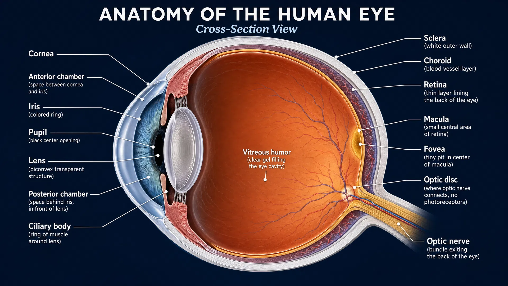
The retinal pigment epithelium, or RPE, is a layer of cells located next to the light-sensing photoreceptors of the retina. Healthy RPE cells have a highly organized, polarized structure. This means that the two sides of each cell have specialized roles, allowing the RPE to support the retina in a coordinated way.
Ciliopathies are a broad group of genetic disorders involving defects in the primary cilium, a tiny cell structure that helps cells sense and respond to signals. Retinal degeneration is a prominent feature of many ciliopathies. The study’s authors note that disease processes may affect both photoreceptors and the RPE during development and maturation.
To investigate shared processes, the team generated RPE cells from induced pluripotent stem cells, or iPSCs. iPSCs are cells that can be made from a person’s cells and then guided in the laboratory to become other cell types, including RPE. These patient-derived iRPE models allow researchers to study disease-associated cell changes in a controlled setting.
A convergent problem across different genes
The researchers examined iRPE made from nine patients with severe retinal degeneration associated with variants in BBS1, BBS10, BBS16, CEP290, LCA5, MYO7A, or PRPF31. These genes are associated with varied disorders and disease severity, yet the cells showed consistent abnormalities.
Across the ciliopathy iRPE models, the researchers found abnormal epithelial polarization. In everyday terms, the RPE cells were not maintaining the organized, specialized state needed for their normal support role in the retina.
They also found poorer mitochondrial health. Mitochondria are often called the cell’s energy-producing structures, but they do much more than generate energy. When mitochondrial health is disrupted, cells may have difficulty sustaining the demanding functions required to maintain tissues such as the retina.
Importantly, the study links these changes to what the authors call mesenchymal drift. Epithelial cells are normally structured and tightly connected to their neighbors. During mesenchymal drift, they begin shifting away from that stable epithelial identity. This shift was driven by dysregulated TGF-β signaling in the patient-derived RPE cells.
The key insight is that this cell-state change appeared despite differences in the underlying disease genes. The study therefore supports the idea that multiple genetic causes of retinal degeneration may converge on shared downstream pathways.
Two potential drug approaches
The investigators identified two drugs as potential candidates for addressing these shared disease features:
- Pioglitazone, described in the study as a mitochondrial metabolic modulator.
- Galunisertib, an inhibitor of TGFBR1, a receptor involved in TGF-β signaling.
These candidates target downstream cellular consequences rather than attempting to replace or edit a specific faulty gene. Galunisertib is particularly relevant to the study’s central finding because it targets the signaling pathway associated with mesenchymal drift. Pioglitazone addresses the accompanying mitochondrial metabolic problem.
This strategy could have an important practical advantage. Single-gene therapies can be highly specific, but developing them for many rare genetic subtypes can be costly and difficult to scale. A treatment aimed at a shared disease mechanism might eventually complement gene-specific approaches and potentially be relevant to several conditions.
However, the study identifies these drugs as potential therapeutic candidates in cell models. The abstract does not establish that either medicine preserves vision, slows retinal degeneration, or is effective in people with Usher syndrome. Further research will need to test how these findings translate in more complex disease models and, ultimately, in carefully designed clinical studies.
Looking ahead: from gene diagnosis to shared biology
For the Usher syndrome community, this research reinforces two connected ideas. First, identifying the causal gene remains crucial, particularly as gene-targeted therapies continue to advance. Second, the genetic diagnosis may not be the only useful treatment guide. Shared cellular pathways may offer additional opportunities for therapy.
The inclusion of MYO7A-derived RPE cells in a broader group of ciliopathy models suggests that researchers can learn from similarities across conditions as well as from their differences. By mapping where diverse genetic disorders converge inside vulnerable retinal cells, studies like this may help expand the treatment landscape—from one-gene-at-a-time strategies toward approaches that protect common cellular functions essential for retinal health.