Why this study matters
Retinitis pigmentosa (RP) is a group of inherited retinal diseases in which light-sensing photoreceptor cells progressively lose function and degenerate. For people and families affected by RP, one of the central challenges is that important changes in the retina may occur before they are easily captured by standard clinical measures. Researchers need sensitive ways to track these changes—both to understand how disease progresses and to evaluate treatments designed to preserve vision.
A 2026 study published in npj biomedical innovations focuses on RP caused by pathogenic variants in the rhodopsin (RHO) gene. Variants in RHO are the most common cause of autosomal dominant RP. The research used adaptive optics (AO) retinal imaging, a technology that can visualize photoreceptors at remarkably high resolution in living eyes, to examine how rod and cone cells are organized across the retina.
The findings suggest that retinal structure can be disrupted in ways that are not fully apparent when looking only at whether cone cells are present or absent. This may be especially important for developing clinical trials aimed at preserving photoreceptors.
The retina’s rods and cones
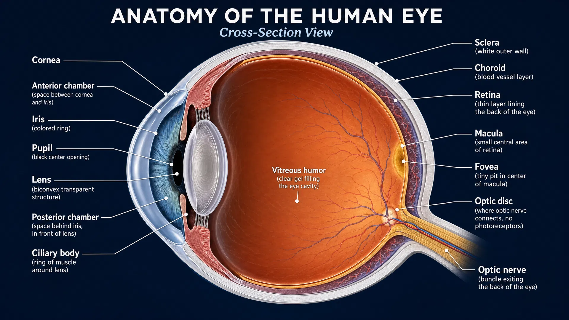
The retina contains two main types of photoreceptors. Rods are especially important for seeing in dim light and for peripheral vision. Cones support central, detailed, and color vision. In RP, rod dysfunction and degeneration are often prominent early features, while cone loss can become increasingly important as disease advances.
Rhodopsin is a light-sensitive protein found in rod photoreceptors. Therefore, it is biologically significant that the study found greater disruption of rod density than cone density at the advancing edge of disease, described by the researchers as the “leading disease front.” This pattern supports the idea that rod damage is a major driver of retinal change in RHO-associated RP.
What adaptive optics revealed
Adaptive optics imaging compensates for optical imperfections in the eye, allowing researchers to inspect the photoreceptor mosaic—the regular cellular arrangement of rods and cones—with far greater detail than is available through conventional retinal imaging.
Using this approach, the investigators identified irregularities in cone organization even in retinal regions that appeared relatively well preserved. In accessible terms, cones may still be present in some areas, but their usual orderly arrangement can already be disturbed. This distinction matters: counting cells alone may not fully describe the health of the photoreceptor layer.
At the leading edge of degeneration, the study found that rod density was disrupted more extensively than cone density. Together, these observations point to a process in which rod degeneration is prominent, while cone structure may become disorganized even before an area appears severely affected.
This is an important refinement in how retinal change may be measured in RHO-related RP. Rather than viewing disease progression only as a gradual disappearance of photoreceptors, researchers may be able to detect earlier alterations in how the remaining cells are spaced and organized.
Why repeated measurements are important
The study also included repeated measurements over time. Longitudinal imaging is essential in inherited retinal disease research because treatment studies need reliable evidence that a change is occurring—or that a treatment is slowing that change.
The researchers concluded that cone-based metrics may be useful for trials seeking to preserve photoreceptor structure. A metric is a measurable feature that can be tracked consistently, such as aspects of cone density or organization. If these measures can detect meaningful structural changes over time, they could become valuable endpoints in studies of potential therapies.
For participants in future trials, sensitive imaging measures may help researchers assess treatment effects before large changes in vision are detectable. They may also help identify which regions of the retina are most informative to follow during a study.
Implications for treatment development
This publication does not test a treatment. Instead, it advances an important part of the treatment-development pathway: identifying precise, repeatable ways to measure retinal preservation.
For RHO-associated RP, therapies may ultimately aim to address the effects of the disease-causing genetic variant, protect vulnerable photoreceptors, or preserve retinal structure for as long as possible. Whatever the therapeutic approach, clinical trials need outcome measures that reflect the biology of the disease. High-resolution AO imaging could help provide that bridge between cellular retinal changes and the evaluation of emerging interventions.
The findings also emphasize the importance of preserving cones, which are critical to useful central vision. Even when cones remain detectable, changes in their organization may signal that the retina is under stress. Measuring those changes could help researchers evaluate whether a therapy is stabilizing the photoreceptor mosaic.
Looking ahead
This research illustrates how advances in imaging are reshaping the study of inherited retinal diseases. In RHO-related RP, adaptive optics can reveal a more detailed picture of rod loss and cone disorganization than broader structural assessments alone.
As longitudinal datasets grow, researchers can further determine which AO-derived cone measures are most consistent, sensitive, and meaningful for clinical trials. The long-term goal is clear: to pair promising treatments with tools capable of showing whether they are preserving the retinal cells that support vision. For families living with RP, this work represents progress not only in seeing the disease more clearly, but also in building stronger foundations for future therapeutic studies.