Why earlier recognition matters
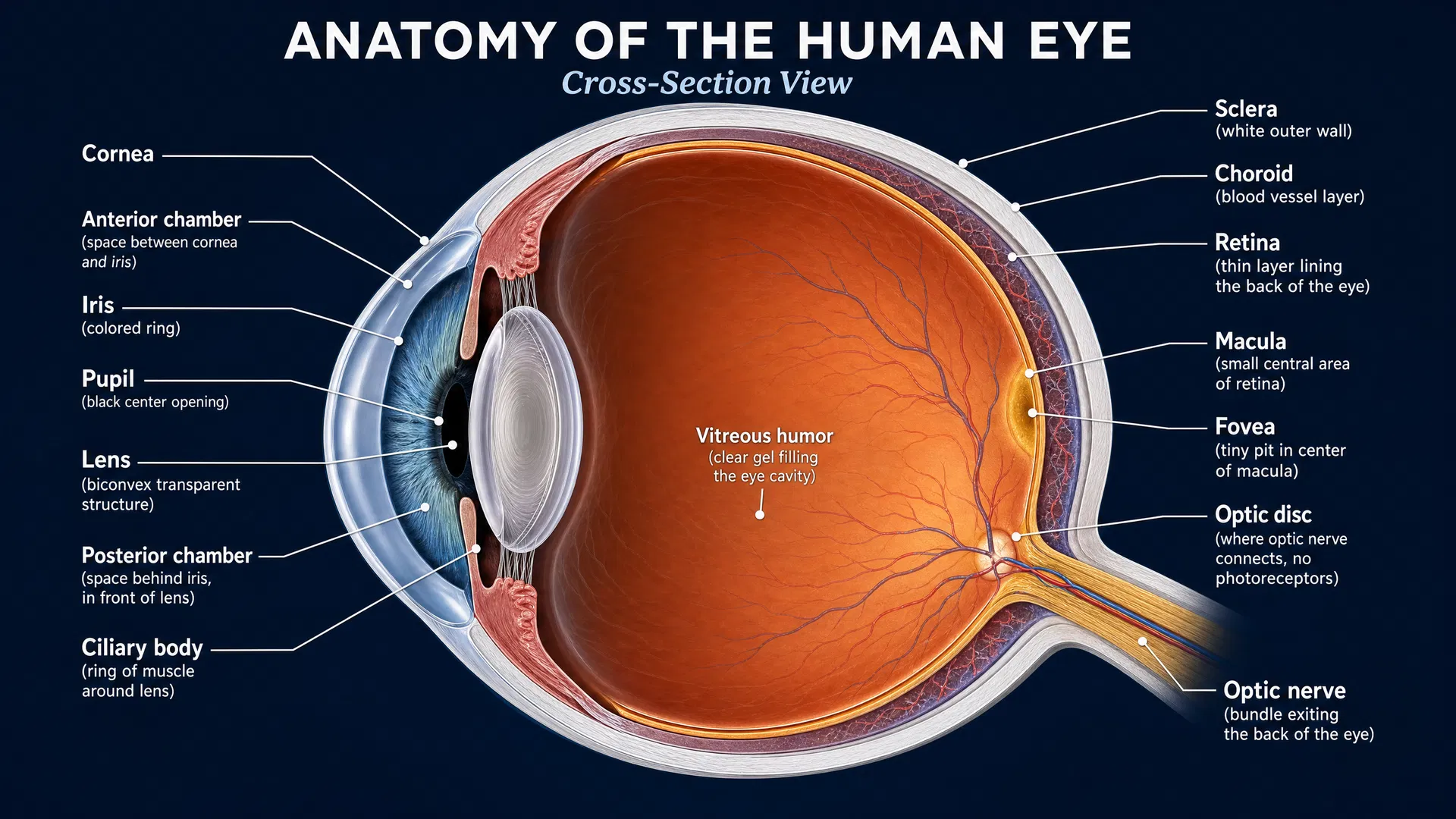
Stickler syndrome (SS) is a rare genetic condition caused by changes in genes that encode collagen, an important structural protein in the body. For many people with Stickler syndrome, eye complications can be among the earliest and most serious features. In particular, the risk of retinal detachment is increased. Because diagnosis is often delayed, ophthalmologic problems may become the first clue that a person has the condition.
This delay can have profound consequences. Retinal detachment can lead to severe visual impairment, including in early childhood. For patients and families, the challenge is not simply recognizing an eye emergency; it is identifying the underlying syndrome soon enough to support appropriate ophthalmologic attention and broader clinical care.
A 2026 study published in Orphanet Journal of Rare Diseases explored a potentially valuable new route to earlier identification: using 2D and 3D facial photographs, combined with supervised machine learning, to recognize facial patterns associated with Stickler syndrome.
Turning facial features into measurable information
Clinicians have long known that some rare genetic conditions can be associated with characteristic facial features. However, visual assessment alone can be subjective, and subtle features may be difficult to recognize—especially when clinicians have limited experience with a rare disorder.
The researchers investigated whether quantitative facial analysis could distinguish people with Stickler syndrome from healthy controls. Rather than relying only on an observer’s impression, this approach uses photographs to capture and analyze measurable aspects of facial shape.
The study used both conventional two-dimensional photographs and three-dimensional facial images. It then applied supervised machine-learning models, including XGBoost and support vector machines (SVM). In simple terms, these computer-based methods learn from labeled examples: images from people known to have Stickler syndrome and images from healthy individuals. The models then look for patterns that help separate the two groups.
Key findings: high accuracy from 2D and 3D images
The findings were striking. Using full-face 2D and 3D photographs, the machine-learning models identified participants with Stickler syndrome with 92% accuracy.
Accuracy improved further when the researchers concentrated on a specific part of the face in 3D images: the orbitonasal region, which includes the area around the eyes and nose. With this focused 3D analysis, classification accuracy reached 96%.
These results suggest that facial morphology—the structure and shape of the face—contains a strong signal that may help identify Stickler syndrome. Importantly, the approach is non-invasive: it relies on photography rather than a surgical procedure or other physically burdensome test.
The orbitonasal finding is also particularly interesting in the context of Stickler syndrome. The study indicates that the area surrounding the eyes and nose may provide especially informative facial measurements for distinguishing affected individuals from healthy controls. This does not mean that one facial feature alone defines the condition. Instead, the algorithm appears to recognize a broader combination of patterns within that region.
What this could mean for patients and clinical care
The central promise of this research is earlier recognition. If facial-image analysis can help flag people who may have Stickler syndrome, it could support referral for further assessment at a time when diagnostic uncertainty remains high.
For families, an earlier pathway to identification could be especially meaningful because of the syndrome’s association with retinal detachment. Recognizing the possibility of Stickler syndrome sooner may help bring ophthalmologic risk into focus before severe visual consequences occur. The study’s authors emphasize that reducing diagnostic delays could help prevent associated complications and improve outcomes.
This type of technology may also be useful in settings where expertise in rare diseases is not immediately available. A quantitative facial-analysis tool could potentially assist clinicians in recognizing when a rare genetic diagnosis should be considered. In this way, artificial intelligence (AI) may help transform photographs into clinically useful screening information.
A screening opportunity, not a replacement for comprehensive diagnosis
The study positions facial morphology as a highly accurate, non-invasive screening tool for rare diseases such as Stickler syndrome. Screening is an important distinction: the goal is to identify individuals who may benefit from further evaluation, not to reduce a complex genetic disorder to a photograph alone.
Stickler syndrome is caused by mutations in collagen-encoding genes, and its clinical significance extends beyond facial appearance. The value of image-based screening lies in its potential to shorten the journey toward recognition and to prompt the appropriate next steps in care—particularly attention to eye health in a condition with increased retinal-detachment risk.
Looking ahead
This research adds to a growing effort to use AI responsibly in rare-disease detection. Its strong performance with both full-face imaging and targeted 3D analysis shows that facial photography may become a useful part of future diagnostic pathways for Stickler syndrome.
The next phase of the research landscape will be translating this promising accuracy into tools that can support real-world clinical decision-making. For the Stickler syndrome community, the potential impact is clear: faster recognition could mean faster access to appropriate evaluation and a greater opportunity to address vision-threatening complications before they cause irreversible harm.