
Why this study matters
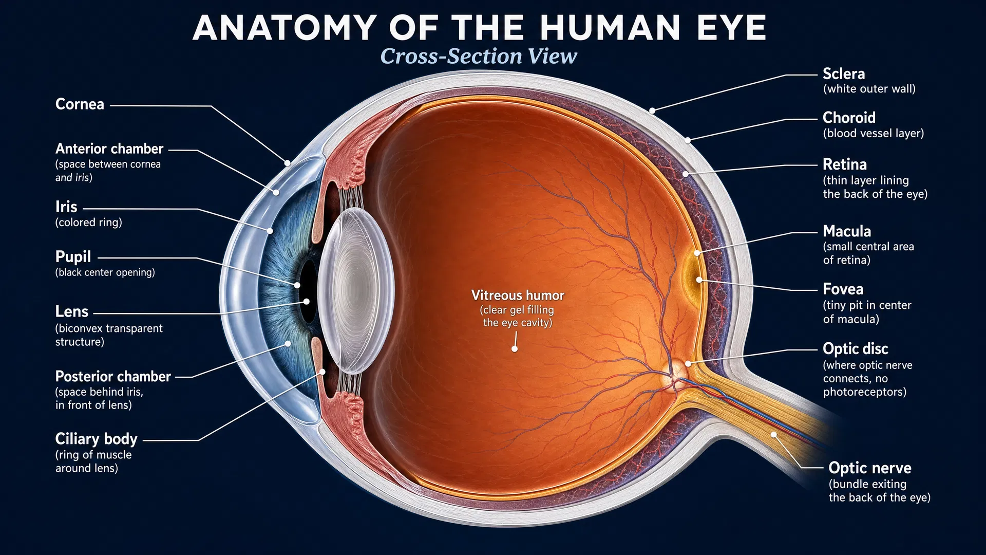
Stargardt disease affects the macula, the central part of the retina responsible for detailed tasks such as reading and recognizing faces. A key goal in Stargardt research is finding reliable ways to measure changes in the retina over time. These measurements could help researchers understand disease progression and, ultimately, assess whether potential treatments are preserving retinal structure.
Adaptive optics flood-illumination ophthalmoscopy (AO-FIO) is an advanced retinal imaging technique that can visualize features of the retina at very high resolution, including cone photoreceptors. Cone cells are critical for sharp central and color vision. Yet obtaining images is only part of the challenge: researchers also need practical, accurate tools to analyze large numbers of images, including images from retinas affected by disease.
A 2026 exploratory study in JMIR Formative Research examined whether GPT-4 could help create an image-analysis script for AO-FIO images from people with Stargardt disease. The work is an early proof of principle—not a validated clinical tool—but it highlights a possible new route for making sophisticated retinal-image analysis more accessible and scalable.
The challenge: analyzing diseased retinas at scale
Dedicated software for AO retinal image analysis is limited, particularly for large-scale measurements and for images from nonhealthy eyes. This is important in Stargardt disease, where changes in the photoreceptor layer may make images more difficult to interpret than images from healthy participants.
The researchers focused on creating an R-language script, meaning a set of computer instructions that could preprocess AO-FIO images and identify image features called “blobs.” In this context, the blobs were intended to support cone detection and quantification. Rather than writing all of the code from scratch, the team used GPT-4 to help generate the script and then refined it through repeated testing.
What the researchers did
The study used AO-FIO images from four initial participants: one healthy individual and three people with Stargardt disease. GPT-4 was prompted to produce R code for image preprocessing and analysis. The resulting code was tested, revised, and retested through trial and error.
After 54 rounds of instructions and refinements, the investigators produced a functional script that identified and quantified blobs in the AO images. A second researcher, who had not been involved in the earlier coding process, then checked the code for errors using images from another four participants—again, one healthy individual and three participants with Stargardt disease.
The investigators also compared cone counts from five small AO-image sections with counts made independently by two human graders and with results from an existing AO-analysis program. Notably, that pre-existing software had been trained using healthy participants. The abstract does not report the level of agreement between the human counts, existing software, and GPT-4-supported script. Instead, the study emphasizes that the new script remains preliminary and nonvalidated.
Key findings in plain language
The central finding was feasibility: GPT-4 could assist researchers in building a working script for a specialized retinal-imaging task. This required substantial human involvement. The model did not simply produce a finished clinical-grade program in one step; researchers iteratively tested the code, identified problems, and fine-tuned it over 54 instruction cycles.
The script was also assessed on images from people with Stargardt disease, not solely on healthy retinal images. That is an important feature of this exploratory work because tools developed only in healthy eyes may not perform as intended when retinal structure is altered by disease.
However, “functional” does not mean “ready for clinical research.” The investigators specifically describe the program as nonvalidated. More refinement, larger-scale testing, and formal validation will be needed before cone counts generated by the script can be relied upon as research measurements.
What this could mean for treatment research
No treatment was tested in this study, and the GPT-4-supported script does not change current care. Its potential relevance lies in measurement. Clinical trials need dependable outcomes that can show whether a treatment may be slowing retinal change or preserving cells. If AO-based cone counts can be rigorously validated, they may become useful biomarkers—measurable indicators that can support studies of disease progression and investigational therapies.
Automated or semi-automated analysis could also reduce the burden of manually counting features in high-resolution images. This may be especially valuable when studies involve many images, repeated visits, and participants with varying degrees of retinal change.
Still, the quality of a biomarker depends on more than the ability to detect features in an image. Researchers will need to determine how well the script performs across a wider range of Stargardt images, how its results compare with expert human assessment, and whether measured cone changes are meaningful for clinical trials.
A promising early step
This study illustrates a practical role for generative AI in retinal research: helping scientists develop specialized analysis tools when dedicated software is scarce. The work also makes clear that expert oversight, testing, and validation remain essential.
For the Stargardt community, the long-term opportunity is better measurement. As imaging methods and analysis tools improve, researchers may be better positioned to track photoreceptor changes and evaluate potential treatments with greater precision. This GPT-4-supported script is not yet a finished answer, but it is an encouraging early experiment in building the tools needed for the next stages of Stargardt disease research.