
What does vision loss look like in Bardet-Biedl syndrome?
Rod-cone dystrophy, from night blindness to a small island of sight. In the words of people who live with it, with the frames from our simulator and the sources behind every statement.

The short answer
Rod-cone dystrophy is the pattern behind retinitis pigmentosa and the eye disease of Bardet-Biedl syndrome: the rods fail first, so the dark and the sides go, then the cones follow, so the center blurs and colors fade. To the child it looks like this in order: a normal daytime world and a difficult night; then things that are not there at the sides, without any sense of a missing piece; then a small, clear island that slowly becomes smaller and softer. Even at the end, most people still see light. Where the pattern comes from a syndrome, it tends to move faster.
- What the person is experiencingNight blindness comes first, usually bringing a child to medical attention in the first decade of life, followed by progressive loss of side vision, fading color discrimination and then loss of sharpness.
- In their wordsThe accounts on our RP page apply here, because the pattern is the same.
- Why it moves faster in a syndromeIn Bardet-Biedl syndrome the retinal disease is one part of a condition that also involves weight, kidneys, hormones and development, and it tends to reach legal blindness in the teens or twenties rather than in middle age.
See it, stage by stage
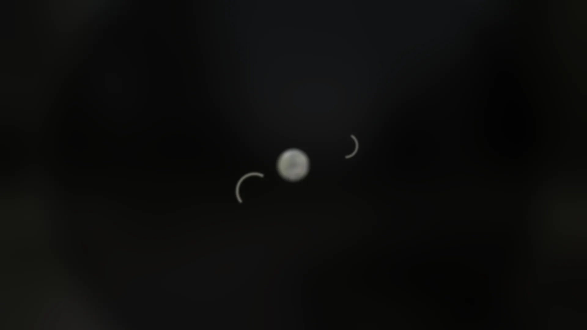
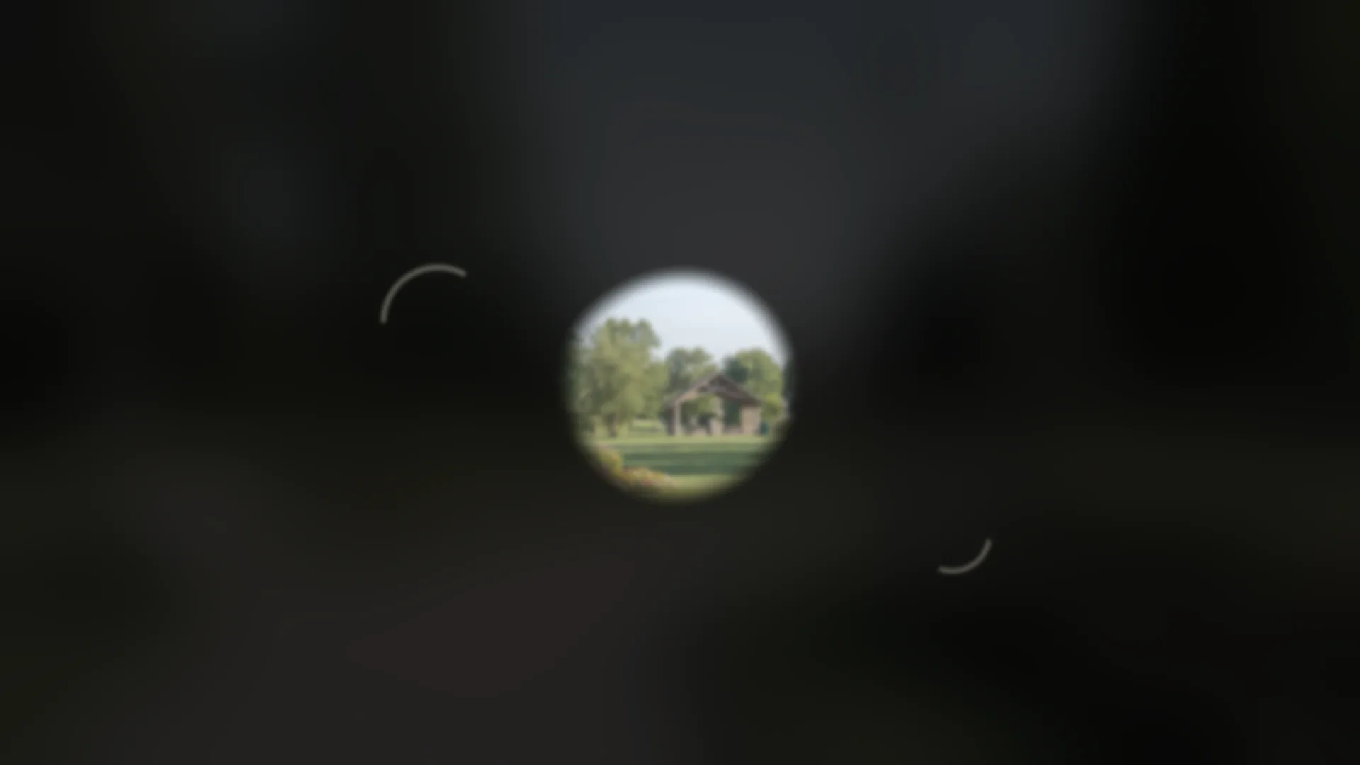
The left view never changes. The right one is the same park as rod-cone dystrophy shows it, drawn from the research our simulator rests on. Pick a stage; the caption tells you what each frame shows and what it cannot.
What the person is experiencing
Night blindness comes first, usually bringing a child to medical attention in the first decade of life, followed by progressive loss of side vision, fading color discrimination and then loss of sharpness 1. In Bardet-Biedl syndrome retinal dystrophy affects 94% of people, and many are legally blind by the second to third decade 1. In non-syndromic RP the timeline is usually slower: many people keep good central vision into their 40s or 50s 2. Even at the end stage, total loss of vision is uncommon; most people retain some light perception 2.
The ring of blind spots in the mid-periphery, 20 to 25 degrees out, widens outward and inward over time; islands of vision may persist for years in the far periphery, and "long after the entire peripheral field is gone, a small oval of intact central field typically remains" 3. Because side-vision loss is "rarely recognized by the affected individual", children may be thought clumsy before anyone measures the field 3.
In their words
The accounts on our RP page apply here, because the pattern is the same. Laura Lawson Visconti, with about 24 degrees of field: "I scan a lot and my brain fills in the information that my eyes miss"; the lost periphery is not black, "I simply do not see anything" 4. Dave Steele on what the tunnel feels like: "It's like a tunnel closing inwards, then you start to lose your central" 5. Hannah, whose cone-rod dystrophy later took her sides, on the brain's cover-up: "your brain does a really great thing where it like makes up everything you can't see" 6.
For the late stage, Hannah describes her field now as "about a pinhole of vision in my left eye" and the loss as "mostly dark. It was mostly like nothing" 6. Helene Lonborg-Moller describes the course of her Usher syndrome as "gradual, changing and progressive. What I could do one year, I could not do the next, loss followed upon loss" 7.
"It's like a tunnel closing inwards, then you start to lose your central."
Why it moves faster in a syndrome
In Bardet-Biedl syndrome the retinal disease is one part of a condition that also involves weight, kidneys, hormones and development, and it tends to reach legal blindness in the teens or twenties rather than in middle age 1. This is why early educational planning is recommended: braille, mobility training, adaptive living skills and computer skills including voice recognition, while making the most of large print while vision remains 1.
What our simulation shows
For the earlier stages, see the RP frames, which share the same model 8.
Signs you might notice in a child
- Fear or hesitation at dusk, asking for lights, reverting to crawling after dark 10.
- Bumping into things, missing balls, tripping over low objects, being called clumsy 3 11.
- Later, holding things close, sitting near the board, and difficulty with colors as the cones become involved 1 12.
- In Bardet-Biedl syndrome, the eye signs usually arrive alongside the other features of the syndrome, and the ERG is more likely to show significant findings after age five 1.
Common questions
Will he be totally blind?
Is the lost area black?
Go deeper
- The rod-cone dystrophy / Bardet-Biedl profileGenes, inheritance, what to expect, and what is in trials.
- For the clinician in the familyNot yet live. The clinician page for this condition is still in review.
- What the other conditions look like
- What does night blindness look like?
- What does retinitis pigmentosa look like?
- What does Stargardt disease look like?
- What does choroideremia look like?
- What does cone dystrophy look like?
- What does achromatopsia look like?
- What does Best disease look like?
- What does Usher syndrome look like?
- What does Leber congenital amaurosis look like?
- Children and familiesSchool, services and support, with early warning signs by age.
References
Sources were read on Oct 22-23, 2026.
- 1.Forsyth RL, Gunay-Aygun M. Bardet-Biedl Syndrome Overview. In: GeneReviews [Internet]. University of Washington, Seattle; 2003, updated 2023. https://www.ncbi.nlm.nih.gov/books/NBK1363/
- 2.O'Neal TB, Tripathy K, Luther EE. Retinitis Pigmentosa. In: StatPearls [Internet]. StatPearls Publishing; 2024. https://www.ncbi.nlm.nih.gov/books/NBK519518/
- 3.GeneReviews [Internet]. Nonsyndromic Retinitis Pigmentosa Overview. University of Washington, Seattle. https://www.ncbi.nlm.nih.gov/books/NBK1417/
- 4.Lawson Visconti L. Going blind in a sighted world (personal blog; first-person account of retinitis pigmentosa). http://lauralawsonvisconti.com/blog/rp
- 5.Hadley. Hadley Presents: The Blind Poet, Dave Steele (podcast transcript; Dave Steele describing retinitis pigmentosa). https://hadleyhelps.org/podcasts/hadley-presents/blind-poet-dave-steele
- 6.Pediatric Retinal Research Foundation. Living with Cone Dystrophy: Hannah's Therapeutic Humor (podcast transcript; first-person account). https://prrf.org/living-with-cone-dystrophy/
- 7.Living with Usher Syndrome: Patient and Physician Perspectives (commentary co-authored by patient Helene Lonborg-Moller with ophthalmologists Yousif Subhi and Line Kessel). 2020. PMC7406578. https://pmc.ncbi.nlm.nih.gov/articles/PMC7406578/
- 8.A Race Against Blindness. How our vision simulations were built: research basis and rendering conventions (Education Hub, Oct 2026). https://araceagainstblindness.org/education/vision-simulations/methodology
- 9.Bittner AK, Diener-West M, Dagnelie G. A survey of photopsias in self-reported retinitis pigmentosa: location of photopsias is related to disease severity. Retina. 2009;29(10):1513-1521. doi:10.1097/IAE.0b013e3181af0d57 https://pmc.ncbi.nlm.nih.gov/articles/PMC2783747/
- 10.Hesperian Health Guides. Disabled Village Children: Causes of Loss of Vision. https://en.hesperian.org/hhg/Disabled_Village_Children:Causes_of_Blindness
- 11.Nemours KidsHealth. Retinitis Pigmentosa (for Parents). https://kidshealth.org/en/parents/retinitis-pigmentosa.html
- 12.Raising Children Network (Australia). Low vision and blindness: babies, children and teenagers. https://raisingchildren.net.au/disability/guide-to-disabilities/assessment-diagnosis/low-vision-blindness-babies-children-teens
- 13.American Academy of Ophthalmology, EyeWiki. Retinitis Pigmentosa. https://eyewiki.aao.org/Retinitis_Pigmentosa
- 14.Gene Vision (UK). Retinitis pigmentosa: for patients. https://gene.vision/knowledge-base/retinitis-pigmentosa-for-patients/
About this page
This page is an additional resource for the community. It is not a substitute for advice from your own eye care team, or for medical journals, textbooks or clinical training. We have done our best to provide accurate and up-to-date information, and the references are listed above, but inaccuracies and omissions may exist. Confirm any medical information with your eye care team or against primary-source medical education resources before relying on it. The simulation frames are approximations built from the cited literature, not a prediction of anyone's vision.